Thursday, March 08, 2007

Epilepsy


Epilepsy : seizure disorder characterized by sudden transient aberration of brain function; associated with motor, sensory, automatic, or psychic disturbances.

A. Seizure : involuntary muscular contraction and disturbances of consciousness from abnormal electrical activity.

B. Risk Factor
1. Brain Injury
2. Infection (meningitis, encephalitis).
3. Water and electrolyte disturbances.
4. Hypoglycemia
5. Tumors
6. Vascular disorders (hypoxia or hypocapnia)


C. Generalized Seizures :


1. Tonic-clonic (grand mal) seizures :
increased excitability of a neuron, possible activation of adjacent neurons, synchronous discharge of impulses, vigorous involuntary sustained muscle spasm (Tonic contractions). Onset of neural fatigue, intermittent muscle spasm (clonic contraction) cessation of muscle spasms, fatigue.

2. Absence (Petit mal) seizures :
unknown etiology, momentary loss of consciousness (10-20 second) usually no recollection of seizures; resumes previously performed action.

3. Minor Motor seizures
a. Myoclonic : involuntary jerking contraction of major muscles; may throw person to the floor.
b. Akinetic : momentary loss of muscle movement.
c. Atonic : total loss for muscle tone; person falls to the floor.

D. Partial (Focal) seizures :
1. Partial motor : arises from region in motor cortex (posterior frontal lobe .
2. Partial sensory : sensory symptoms occur with partial seizures activity; varices with region in brain; transient.
3. Partial complex (psychomotor) : arises out of anterior temporal lobe; frequently begins with an aura; characteristic feature is automatism (lip smacking, chewing, patting body, picking at clothes); lasts from 2-3 minutes to 15 minutes do not restrain.

Read more here............

Tuesday, December 26, 2006

“Hypertension”


Definition

Hypertension, commonly referred to as "high blood pressure", is a medical condition where the blood pressure is chronically elevated. While it is formally called arterial hypertension, the word "hypertension" without a qualifier usually refers to arterial hypertension. Persistent hypertension is one of the risk factors for strokes, heart attacks, heart failure and arterial aneurysm, and is a leading cause of chronic renal failure.

Hypertension can be classified as either essential or secondary. Essential hypertension is the term used when no specific medical cause can be found to explain a patient's condition. Secondary hypertension means that the high blood pressure is a result of (i.e. secondary to) another condition, such as kidney disease or certain tumours.


Recently, the JNC 7 (The Seventh Report of the Joint National Committee on Prevention, Detection, Evaluation, and Treatment of High Blood Pressure) has defined blood pressure 120/80 mmHg to 139/89 mmHg as "prehypertension." Prehypertension is not a disease category; rather, it is a designation chosen to identify individuals at high risk of developing hypertension.

The Mayo Clinic website indicates that your blood pressure is "normal if it's below 120/80" but that "some data indicate that 115/75 mm Hg should be the gold standard."

"In patients with diabetes mellitus or kidney disease studies have shown that blood pressure over 130/80 mmHg should be considered a risk factor and warrants treatment. Even lower numbers are considered diagnostic using home blood pressure monitoring devices. Contents

Hypertension means high blood pressure. This generally means:
Systolic blood pressure is consistently over 140 (systolic is the "top" number of your blood pressure measurement, which represents the pressure generated when the heart beats). Diastolic blood pressure is consistently over 90 (diastolic is the "bottom" number of your blood pressure measurement, which represents the pressure in the vessels when the heart is at rest). Either or both of these numbers may be too high.

Pre-hypertension is when your systolic blood pressure is between 120 and 139 or your diastolic blood pressure is between 80 and 89 on multiple readings. If you have pre-hypertension, you are likely to develop high blood pressure at some point. Therefore, your doctor will recommend lifestyle changes to bring your blood pressure down to normal range.

Etiology of Essential Hypertension

1. Environment
A number of environmental factors have been implicated in the development of hypertension, including salt intake, obesity, occupation, alcohol intake, family size, excessive noise exposure,[2] and crowding.

2. Salt Sensitivity
Sodium is the environmental factor that has received the greatest attention. It is to be noted that approximately 60% of the essential hypertension population is responsive to sodium intake.

3. Role of Rennin
Rennin is an enzyme secreted by the juxtaglomerular cells of the kidney and linked with aldosterone in a negative feedback loop.The range of plasma rennin activities observed in hypertensive subjects is broader than in normotensive individuals. In consequence, some hypertensive patients have been defined as having low-rennin and others as having high-rennin essential hypertension.

4. Insulin Resistance
Insulin is a polypeptide hormone secreted by the pancreas. Its main purpose is to regulate the levels of glucose in the body, it also has some other effects. Insulin resistance and/or hyperinsulinemia have been suggested as being responsible for the increased arterial pressure in some patients with hypertension. This feature is now widely recognized as part of syndrome X, or the metabolic syndrome.

5. Sleep Apnoea
Sleep apnoea is a common, under recognized cause of hypertension. It is best treated with weight loss and nocturnal nasal positive airway pressure.

6.Genetics
Hypertension is one of the most common complex genetic disorders, with genetic heritability averaging 30%. Data supporting this view emerge from animal studies as well as in population studies in humans. Most of these studies support the concept that the inheritance is probably multifactor or that a number of different genetic defects each have an elevated blood pressure as one of their phenotypic expressions.

More than 50 genes have been examined in association studies with hypertension, and the number is constantly growing.

7. Other Etiologies
There are some anecdotal or transient causes of high blood pressure. These are not to be confused with the disease called hypertension in which there is an intrinsic physiopathological mechanism as described above.

Etiology of Secondary Hypertension

Only in a small minority of patients with elevated arterial pressure can a specific cause be identified. These individuals will probably have an endocrine or renal defect that if corrected would bring blood pressure back to normal values.

1. Renal Hypertension
Hypertension produced by diseases of the kidney. A simple explanation for renal vascular hypertension is that decreased perfusion of renal tissue due to stenosis of a main or branch renal artery activates the renin-angiotensin system.

2. Adrenal Hypertension
Hypertension is a feature of a variety of adrenal cortical abnormalities. In primary aldosteronism there is a clear relationship between the aldosterone-induced sodium retention and the hypertension.
In patients with pheochromocytoma increased secretion of catecholamines such as epinephrine and norepinephrine by a tumor (most often located in the adrenal medulla) causes excessive stimulation of [adrenergic receptors], which results in peripheral vasoconstriction and cardiac stimulation. This diagnosis is confirmed by demonstrating increased urinary excretion of epinephrine and norepinephrine and/or their metabolites (vanillylmandelic acid).

3. Hypercalcemia

4. Coarctation of the Aorta

5. Diet

Certain medications, especially NSAIDS (Motrin/ibupofen) and steroids can cause hypertension. Ingestion of imported licorice (Glycyrrhiza glabra) can cause secondary hypoaldosteronism, which itself is a cause of hypertension.
Age. Over time, the number of collagen fibers in artery and arteriole walls increases, making blood vessels stiffer. With the reduced elasticity comes a smaller cross-sectional area in systole, and so a raised mean arterial blood pressure.

Pathophysiology

Most of the secondary mechanisms associated with hypertension are generally fully understood, and are outlined at secondary hypertension. However, those associated with essential (primary) hypertension are far less understood. What is known is that cardiac output is raised early in the disease course, with total peripheral resistance (TPR) normal; over time cardiac output drops to normal levels but TPR is increased. Three theories have been proposed to explain this:
Inability of the kidneys to excrete sodium, resulting in natriuretic factors such as Atrial Natriuretic Factor being secreted to promote salt excretion with the side-effect of raising total peripheral resistance.
An overactive renin / angiotension system leads to vasoconstriction and retention of sodium and water. The increase in blood volume leads to hypertension.
An overactive sympathetic nervous system, leading to increased stress responses.

Is is also known that hypertension is highly heritable and polygenic (caused by more than one gene) and a few candidate genes have been postulated in the etiology of this condition

Signs and symptoms

Hypertension is usually found incidentally - "case finding" - by healthcare professionals. It normally produces no symptoms. Malignant hypertension (or accelerated hypertension) is distinct as a late phase in the condition, and may present with headaches, blurred vision and end-organ damage. It is recognised that stressful situations can increase the blood pressure;

Hypertension is often confused with mental tension, stress and anxiety. While chronic anxiety is associated with poor outcomes in people with hypertension, it alone does not cause it.

Hypertensive urgencies and emergencies
Hypertension is rarely severe enough to cause symptoms. These typically only surface with a systolic blood pressure over 240 mmHg and/or a diastolic blood pressure over 120 mmHg. These pressures without signs of end-organ damage (such as renal failure) are termed "accelerated" hypertension. When end-organ damage is possible or already ongoing, but in absence of raised intracranial pressure, it is called hypertensive emergency. Hypertension under this circumstance needs to be controlled, but prolonged hospitalization is not necessarily required. When hypertension causes increased intracranial pressure, it is called malignant hypertension. Increased intracranial pressure causes papilledema, which is visible on ophthalmoscopic examination of the retina.

Complications

While elevated blood pressure alone is not an illness, it often requires treatment due to its short- and long-term effects on many organs. The risk is increased for:
Cerebrovascular accident (CVAs or strokes)
Myocardial infarction (heart attack)
Hypertensive cardiomyopathy (heart failure due to chronically high blood pressure)
Hypertensive retinopathy - damage to the retina
Hypertensive nephropathy - chronic renal failure due to chronically high blood pressure

Pregnancy
Main article: Hypertension of pregnancy

Although few women of childbearing age have high blood pressure, up to 10% develop hypertension of pregnancy. While generally benign, it may herald three complications of pregnancy: pre-eclampsia, HELLP syndrome and eclampsia. Follow-up and control with medication is therefore often necessary.

Diagnosis

1. Measuring blood pressure
Diagnosis of hypertension is generally on the basis of a persistently high blood pressure. Usually this requires three separate measurements at least one week apart. Exceptionally, if the elevation is extreme, or end-organ damage is present then the diagnosis may be applied and treatment commenced immediately.

Obtaining reliable blood pressure measurements relies on following several rules and understanding the many factors that influence blood pressure reading. For instance, measurements in control of hypertension should be at least 1 hour after caffeine, 30 minutes after smoking and without any stress. Cuff size is also important. The bladder should encircle and cover two-thirds of the length of the arm. The patient should be sitting for a minimum of five minutes. The patient should not be on any adrenergic stimulants, such as those found in many cold medications.

When taking manual measurements, the person taking the measurement should be careful to inflate the cuff suitably above anticipated systolic pressure. A stethoscope should be placed lightly over the brachial artery. The cuff should be at the level of the heart and the cuff should be deflated at a rate of 2 to 3 mmHg/s. Systolic pressure is the pressure reading at the onset of the sounds described by Korotkoff (Phase one). Diastolic pressure is then recorded as the pressure at which the sounds disappear (K5) or sometimes the K4 point, where the sound is abruptly muffled. Two measurements should be made at least 5 minutes apart, and, if there is a discrepancy of more than 5 mmHg, a third reading should be done. The readings should then be averaged. An initial measurement should include both arms. In elderly patients who particularly when treated may show orthostatic hypotension, measuring lying sitting and standing BP may be useful. The BP should at some time have been measured in each arm, and the higher pressure arm preferred for subsequent measurements.

BP varies with time of day, as may the effectiveness of treatment, and archetypes used to record the data should include the time taken. Analysis of this is rare at present. Automated machines are commonly used and reduce the variability in manually collected readings. Routine measurements done in medical offices of patients with known hypertension may incorrectly diagnose 20% of patients with uncontrolled hypertension .


Distinguishing primary vs. secondary hypertension

Once the diagnosis of hypertension has been made it is important to attempt to exclude or identify reversible (secondary) causes.
Over 90% of adult hypertension has no clear cause and is therefore called essential/primary hypertension. Often, it is part of the metabolic "syndrome X" in patients with insulin resistance: it occurs in combination with diabetes mellitus (type 2), combined hyperlipidemia and central obesity.
In hypertensive children most cases are secondary hypertension, and the cause should be pursued diligently.

Investigations commonly performed in newly diagnosed hypertension

Tests are undertaken to identify possible causes of secondary hypertension, and seek evidence for end-organ damage to the heart itself or the eyes (retina) and kidneys. Diabetes and raised cholesterol levels being additional risk factors for the development of cardiovascular disease are also tested for as they will also require management.

Blood tests commonly performed include:
Creatinine (renal function) - to identify both underlying renal disease as a cause of hypertension and conversely hypertension causing onset of kidney damage. Also a baseline for later monitoring the possible side-effects of certain antihypertensive drugs.
Electrolytes (sodium, potassium)
Glucose - to identify diabetes mellitus
Cholesterol

Additional tests often include:
Testing of urine samples for proteinuria - again to pick up underlying kidney disease or evidence of hypertensive renal damage.
Electrocardiogram (EKG/ECG) - for evidence of the heart being under strain from working against a high blood pressure. Also may show resulting thickening of the heart muscle (left ventricular hypertrophy) or of the occurrence of previous silent cardiac disease (either subtle electrical conduction disruption or even a myocardial infarction).
Chest X-ray - again for signs of cardiac enlargement or evidence of cardiac failure.

Epidemiology

The level of blood pressure regarded as deleterious has been revised down during years of epidemiological studies. A widely quoted and important series of such studies is the Framingham Heart Study carried out in an American town: Framingham, Massachusetts. The results from Framingham and of similar work in Busselton, Western Australia have been widely applied. To the extent that people are similar this seems reasonable, but there are known to be genetic variations in the most effective drugs for particular sub-populations. Recently (2004), the Framingham figures have been found to overestimate risks for the UK population considerably. The reasons are unclear. Nevertheless the Framingham work has been an important element of UK health policy.

Treatment

1. Lifestyle modification
Doctors recommend weight loss and regular exercise as the first steps in treating mild to moderate hypertension. These steps are highly effective in reducing blood pressure, although most patients with moderate or severe hypertension end up requiring indefinite drug therapy to bring their blood pressure down to a safe level. Discontinuing smoking does not directly reduce blood pressure, but is very important for people with hypertension because it reduces the risk of many dangerous outcomes of hypertension, such as stroke and heart attack.

Mild hypertension is usually treated by diet, exercise and improved physical fitness. A diet rich in fruits and vegetables and low fat or fat-free dairy foods and moderate or low in sodium lowers blood pressure in people with hypertension. This diet is known as the DASH diet (Dietary Approaches to Stop Hypertension), and is based on National Institutes of Health sponsored research. Dietary sodium (salt) may worsen hypertension in some people and reducing salt intake decreases blood pressure in a third of people. Regular mild exercise improves blood flow, and helps to lower blood pressure. In addition, fruits, vegetables, and nuts have the added benefit of increasing dietary Potassium, which offsets the effect of sodium and acts on the kidney to decrease blood pressure.

Reduction of environmental stressors such as high sound levels and over-illumination can be an additional method of ameliorating hypertension. Biofeedback is also used particularly device guided paced breathing

2. Medications

a. Antihypertensives

There are many classes of medications for treating hypertension, together called antihypertensives, which — by varying means — act by lowering blood pressure. Evidence suggests that reduction of the blood pressure by 5-6 mmHg can decrease the risk of stroke by 40%, of coronary heart disease by 15-20%, and reduces the likelihood of dementia, heart failure, and mortality from vascular disease.

The aim of treatment should be blood pressure control to <140/90 mmHg for most patients, and lower in certain contexts such as diabetes or kidney disease (some medical professionals recommend keeping levels below 120/80 mmHg)[4]. Each added drug may reduce the systolic blood pressure by 5-10 mmHg, so often multiple drugs are necessary to achieve blood pressure control.

Commonly used drugs include:
ACE inhibitors such as captopril, enalapril, fosinopril (Monopril®), lisinopril (Zestril®), quinapril, ramipril (Altace®)
Angiotensin II receptor antagonists: eg, irbesartan (Avapro®), losartan (Cozaar®), valsartan (Diovan®), candesartan (Atacand®)
Alpha blockers such as doxazosin, prazosin, or terazosin
Beta blockers such as atenolol, labetalol, metoprolol (Lopressor®, Toprol-XL®)
Calcium channel blockers such as amlodipine (Norvasc®), diltiazem, verapamil
Diuretics: eg, bendroflumethiazide, chlortalidone, hydrochlorothiazide
(also called HCTZ)
Finally combination products (which usually contain HCTZ and one other drug)

Which type of many medications should be used initially for hypertension has been the subject of several large studies and various national guidlelines.

The ALLHAT study showed a slightly better outcome and cost-effectiveness for the thiazide diuretic chlortalidone compared to anti-hypertensives. Whilst a subsequent smaller study (ANBP2) did not show this small difference in outcome and actually showed a slightly better outcome for ACE-inhibitors in older male patients.[10]

Whilst thiazides are cheap, effective, and recommended as the best first-line drug for hypertension by many experts, they are not prescribed as often as some newer drugs. Arguably, this is because they are off-patent and thus rarely promoted by the drug industry.[11] Although physicians may start with non-thiazide antihypertensive medications if there is a compelling reason to do so. An example is the use of ACE-inhibitors in diabetic patients who have evidence of kidney disease, as they have been shown to both reduce blood pressure and slow the progression of diabetic nephropathy.[12] In patients with coronary artery disease or a history of a heart attack, beta blockers and ACE-inhibitors both lower blood pressure and protect heart muscle over a lifetime, leading to reduced mortality.

References :

Wang Y, Wang QJ. The prevalence of prehypertension and hypertension among US adults according to the new joint national committee guidelines: new challenges of the old problem. Arch Intern Med. 2004;164(19):2126-34.

Seventh Report of the Joint National Committee on Prevention, Detection, Evaluation, and Treatment of High Blood Pressure (JNC 7). Rockville, Md. National Heart, Lung, and Blood Institute, US Department of Health and Human Services; August 2004. National Institutes of Health Publication No. 04-5230.

Eyre H, Kahn R, Robertson RM, et al. Preventing cancer, cardiovascular disease, and diabetes: A common agenda for the American Cancer Society, the American Diabetes Association, and the American Heart Association. Circulation. 2004;109(25):3244-55.

Whelton PK, He J, Appel LJ, et al. Primary prevention of hypertension: Clinical and public health advisory from The National High Blood Pressure Education Program. JAMA. 2002;288(15):1882-8.

Read more here............

Tuesday, April 25, 2006

"Intravenous Therapy"


Intravenous therapy or IV therapy is the administration of liquid substances directly into a vein. It can be intermittent or continuous; continuous administration is called an intravenous drip. The word intravenous simply means "within a vein", but is most commonly used to refer to IV therapy.

A. Indications :
1. Establish or maintain a fluid or electrolyte balance
2. Administer continuous or intermittent medication
3. Administer bolus medication
4. Administer fluid to keep vein open (KVO)
5. Administer blood or blood components
6. Administer intravenous anesthetics
7. Maintain or correct a patient's nutritional state
8. Administer diagnostic reagents
9. Monitor hemodynamic functions

B. IV Devices :
a. Steel Needles :
Example: Butterfly catheter. They are named after the wing-like plastic tabs at the base of the needle. They are used to deliver small quantities of medicines, to deliver fluids via the scalp veins in infants, and sometimes to draw blood samples (although not routinely, since the small diameter may damage blood cells). These are small gauge needles

b. Over the Needle Catheters
Example: peripheral IV catheter. This is the kind of catheter you will primarily be using. Also see the close up view of the catheter/needle tip in the next section ("inside the needle catheters").

Catheters (and needles) are sized by their diameter, which is called the gauge. The smaller the diameter, the larger the gauge. Therefore, a 22-gauge catheter is smaller than a 14-gauge catheter. Obviously, the greater the diameter, the more fluid can be delivered. To deliver large amounts of fluid, you should select a large vein and use a 14 or 16-gauge catheter. To administer medications, an 18 or 20-gauge catheter in a smaller vein will do.

C. IV Fluids :
Intravenous fluids are usually provided to:
- Provide volume replacement
- Administer medications, including electrolytes
- Monitor cardiac functions

For example, a patient comes into the ED with gastroenteritis and is dehydrated from vomiting and diarrhea. Acutely, she receives a fluid bolus to expand her intravascular volume. Her blood chemistry shows that her electrolytes are a bit off, so the IV fluid is adjusted to bring them within normal parameters. She is also given medication for nausea via her IV. She will remain on maintenance IV fluids until she is able to drink adequate amounts of fluids.

There are three main types of fluids:
- Isotonic fluids:
Can be helpful in hypotensive or hypovolemic patients.
Can be harmful. There is a risk of fluid overloading, especially in patients with CHF and hypertension.
Examples: Lactated Ringer's (LR), NS (normal saline, or 0.9% saline in water

- Hypotonic fluids:
Can be helpful when cells are dehydrated such as a dialysis patient on diuretic therapy. May also be used for hyperglycemic conditions like diabetic ketoacidosis, in which high serum glucose levels draw fluid out of the cells and into the vascular and interstitial compartments.
Can be dangerous to use because of the sudden fluid shift from the intravascular space to the cells. This can cause cardiovascular collapse and increased intracranial pressure (ICP) in some patients.
Example: .45% NaCl, 2.5% dextrose

- Hypertonic fluids :
Can help stabilize blood pressure, increase urine output, and reduce edema.
Rarely used in the prehospital setting. Care must be taken with their use. Dangerous in the setting of cell dehydration.
Examples: D5% .45% NaCl, D5% LR, D5% NS, blood products, and albumin.

Flow Rates :
You will often need to calculate IV flow rates. The administration sets come in two basic sizes:
1.Microdrip sets, Allow 60 drops (gtts) / mL through a small needle into the drip
chamber (Good for medication administration or pediatric fluid delivery).
2.Macrodrip sets, Allow 10 to 15 drops / mL into the drip chamber (Great for rapid
fluid delivery. Also used for routine fluid delivery).
3.Fluid may be ordered at a KVO rate. This means to Keep the Vein Open, or run in
fluids very slowly, enough to keep the vein open, but not really deliver much
volume.At times, you may desire a faster flow rate. This is usually expressed in
mLs / hour. In other words, how much fluid do you want your patient to receive
each hour? A common "maintenance" amount, for instance, would be "run it in at 125
an hour". Your patient would receive 125 mL of fluid every hour.

This is usually done by counting the number of drops that fall into the clear drip chamber on the IV administration set in one minute. To do this, you must know what size administration set you are using (micro or macrodrip). Plug the numbers into the following formula and you've got it!
(volume in mL) x (drip set) gtts
------------------------------------ = ------
(time in minutes) min

D. Vein Selection:
Veins of the Hand
1. Digital Dorsal veins
2. Dorsal Metacarpal veins
3. Dorsal venous network
4. Cephalic vein
5. Basilic vein


Veins of the Forearm
1. Cephalic vein
2. Median Cubital vein
3. Accessory Cephalic vein
4. Basilic vein
5. Cephalic vein
6. Median antebrachial vein

E. Technique:
Remember the four rights:Do I have the right patient?Do I have the right solution?Do I have the right drug?Do I have the right route?.
Preparation

It is important to gather all the necessary supplies before you begin. You will need: Absorbent disposable sheet, 1 alcohol prep pad, 1 betadine swab, Tourniquet, IV catheter, IV tubing, Bag of IV fluid. 4 pieces of tape (preferably paper tape or easy to remove tape which has been precut to approximately 4 inches (10cm) in length and taped conveniently to the table or stretcher. Disposable gloves, Gauze (several pieces of 4x4 or 2x2)

Prepare the IV fluid administration set.Inspect the fluid bag to be certain it contains the desired fluid, the fluid is clear, the bag is not leaking, and the bag is not expired.
Select either a mini or macro drip administration set and uncoil the tubing. Do not let the ends of the tubing become contaminated.Close the flow regulator (roll the wheel away from the end you will attach to the fluid bag).Remove the protective covering from the port of the fluid bag and the protective covering from the spike of the administration set.Insert the spike of the administration set into the port of the fluid bag with a quick twist. Do this carefully. Be especially careful to not puncture yourself!

Hold the fluid bag higher than the drip chamber of the administration set. Squeeze the drip chamber once or twice to start the flow. Fill the drip chamber to the marker line (approximately one-third full). If you overfill the chamber, lower the bag below the level of the drip chamber and squeeze some fluid back into the fluid bag. Open the flow regulator and allow the fluid to flush all the air from the tubing. Let it run into a trash can or even the (now empty) wrapper the fluid bag came in. You may need to loosen or remove the cap at the end of the tubing to get the fluid to flow although most sets now allow flow without removal. Take care not to let the tip of the administration set become contaminated.

Turn off the flow and place the sterile cap back on the end of the administration set (if you've had to remove it). Place this end nearby so you can reach it when you are ready to connect it to the IV catheter in the patient's vein.
Perform the venipuncture
Be sure you have introduced yourself to your patient and explained the procedure. Apply a tourniquet high on the upper arm. It should be tight enough to visibly indent the skin, but not cause the patient discomfort. Have the patient make a fist several times in order to maximize venous engorgement. Lower the arm to increase vein engorgement.
Select the appropriate vein. If you cannot easily see a suitable vein, you can sometimes feel them by palpating the arm using your fingers (not your thumb) The vein will feel like an elastic tube that "gives" under pressure. Tapping on the veins, by gently "slapping" them with the pads of two or three fingers may help dilate them. If you still cannot find any veins, then it might be helpful to cover the arm in a warm, moist compress to help with peripheral vasodilatation. If after a meticulous search no veins are found, then release the tourniquet from above the elbow and place it around the forearm and search in the distal forearm, wrist and hand. If still no suitable veins are found, then you will have to move to the other arm. Be careful to stay away from arteries, which are pulsatile.


Don disposable gloves. Clean the entry site carefully with the alcohol prep pad. Allow it to dry. Then use a betadine swab. Allow it to dry. Use both in a circular motion starting with the entry site and extending outward about 2 inches. (Using alcohol after betadine will negate the effect of the betadine) Note that some facilities may require an alcohol prep without betadine or sometimes alcohol after betadine. Go with the rules for your facility.

To puncture the vein, hold the catheter in your dominant hand. With the bevel up, enter the skin at about a 30 degree angle and in the direction of the vein. Use a quick, short, jabbing motion. After entering the skin, reduce the angle of the catheter until it is nearly parallel to the skin. If the vein appears to "roll" (move around freely under the skin), begin your venipuncture by apply counter tension against the skin just below the entry site using your nondominant hand. Many people use their thumb for this. Pull the skin distally toward the wrist in the opposite direction the needle will be advancing. Be carefully not to press too hard which will compress blood flow in the vein and cause the vein to collapse. Then pierce the skin and enter the vein as above.
Advance the catheter to enter the vein until blood is seen in the "flash chamber" of the catheter.

If not successful
If you are unsuccessful in entering the vein and there is no flashback, then slowly withdraw the catheter, without pulling all the way out, and carefully watch for the flashback to occur. If you are still not within the vein, then advance it again in a 2nd attempt to enter the vein. While withdrawing always stop before pulling all the way out to avoid repeating the painful initial skin puncture. If after several manipulations the vein is not entered, then release the tourniquet, place a gauze over the skin puncture site, withdraw the catheter and tape down the gauze. Try again in the other arm.
Otherwise,After entering the vein, advance the plastic catheter (which is over the needle) on into the vein while leaving the needle stationary. The hub of the catheter should be all the way to the skin puncture site. The plastic catheter should slide forward easily. Do not force it!!

(release the tourniquet)
Apply gentle pressure over the vein just proximal to the entry site to prevent blood flow. Remove the needle from within the plastic catheter. Dispose of the needle in an appropriate sharps container. NEVER reinsert the needle into the plastic catheter while it is in the patient's arm! Reinserting the needle can shear off the tip of the plastic catheter causing an embolus. Remove the protective cap from the end of the administration set and connect it to the plastic catheter. Adjust the flow rate as desired.

Tape the catheter in place using the strips of tape and a sterile 2X2 or a clear dressing. It is advisable not to use the "chevron" taping technique.
Label the IV site with the date, time, and your initials.Monitor the infusion for proper flow into the vein (in other words, watch for infiltration).

Occasionally, you may inadvertently enter an artery. You'll recognize this because bright red blood is quickly seen in the IV tubing and the IV bag because of the high pressure that exists. If this occurs, stop the fluid flow, remove the catheter, and put pressure on the site for at least 5 minutes.

To discontinue an IV
Remember to observe universal precautions. Start by clamping off the flow of fluids. Then gently peel the tape back toward the IV site. As you get closer to the site and the catheter, stabilize the catheter and remove the rest of the tape from the patient's skin. Then place a 4 x 4 gauze over the site and gently slide the plastic catheter out of the patient's arm. Use direct pressure for a few minutes to control any bleeding. Finally, place a band aide over the site.

Complications: Bruising, Cellulitis, Infiltrate, Extravasation, Phlebitis, Systemic Complications.


# Refences :
Steve Martin, PhD(c), PA-C
Nova Southeastern University PA Program

Read more here............

"Diverticulosis"


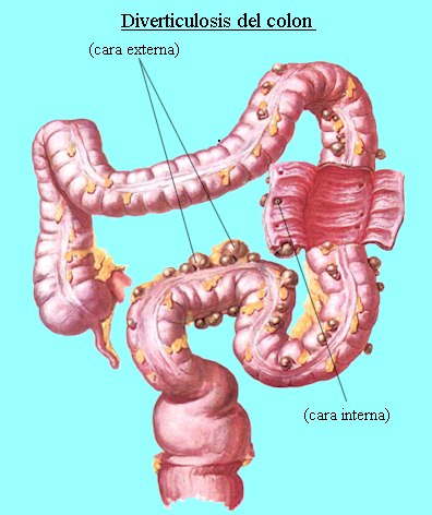
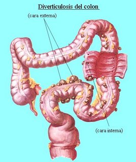
Deverticulosis is the condition in which an individual has multiple diverticulae. A Diverticulum is a douch or sac in the walls of a canal or an organ.
Diverticulosis usually occurs in about 10 % of individuals over 40 years of age and nearly 50 % of person over age 60; only small percentage develop diverticulitis.
The condition is most common in sigmoid colon. Small bowel diverticula are unsual, but when they occur, they are multiple. They may act as areas of statis and bacterial overgrowth, leading to malabsorption of fat and vitamin B12.
If the diverticulum perforates, local abcess or peritonitis results.

Predisposing Factors :
a. Probable congenital predisposition.
b. Weakening and degeneration of muscular wall of the intestine, causing herniation of the lining of the mucous membrane through a muscle.
c. Chronic overdistention of the large bowel.
d. Diet low in roughage-reduce and fecal residue, narrows the bowel lumenand leads to higher pressure intra abdominally during defecation.

Specific Clinical Signs
a. Diverticulosis :
- Bowel irregularity
- Constipation
- Diarrhea
- Sudden massive hemorrhage
b. Milder form of Diverticulitis :
- Mild lower abdominal cramps
- Bowel irregularity, constipation and diarrhea
- Mild nausea, gas, low grade fever and leukocytosis
c. Moderatly severa acute Diverticulitis :
- Crampy pain in lower left quadrant of abdomen
- Low grade fever, chill and leukocytosis
- Ruptured diverticulum produces abscess, ruptured diverticulum near a blood vessel may cause massive hommorhage.
d. Chronic diverticulitis may cause adhesions that narrow the bowels opening and can partial or complete bowel obstruction.


DIAGNOSTIC EVALUATION
1. History, physical examination, laboratory evaluation
2. Flat film of abdomen
3. Ultrasonography, CT-Scan
4. Sigmoidoscopy; possibly colonoscopy
5. Barium Enema (After Infection Subsides)

Surgical Management :
If there is little response to medical treatment or if complications such as hemorrhage, obstructions or perforation occurs, surgery is necessary.
Hemorrhage may require transfusion if hemorrhage is life threatening, total collection is with ileostomy and preservation of rectal stump may be required. Continuity of the bowel is reestablished subsequently.
Intestinal obstruction /perforations-temporary colostomy is sometimes performed to divert fecal stream.

Nursing Care Analysis :
1.a. Nursing Diagnosis
Pain Related to intestinal discomfort, diarrhea and/or constipation

b. Objectives
Patient will have tolerable pain/absence of pain

c.Nursing Intervention
· Assess pts condition
· Observe for sign and location of pain, type and severity
· Auscultative for bowel sounds
· Palpate abdomen to determine rigidity or tenderness due to perforation or peritonitis
· Administer analgesia as prescribed or anti cholinergic as prescribed to decrease colon spasm.

d.Evaluation
Patient expressed relief of pain and has a decrease in symptoms.

2. a.Nursing Diagnosis
Altered nutrition less than body requirements related to diarrhea, fluid and electrolyte loss, nausea and vomiting

b. Objectives
To maintain adequate nutrition· Follow prescribed diet that is high in fat residue low in sugar

c. Nursing Intervention
· Encourage increase fluid intake
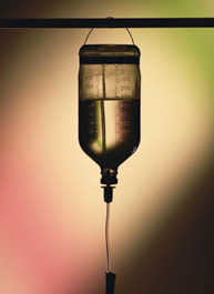
· Regulate IVF properly in case of NPO
· Monitor intake output

d. Evaluation
Consumes prescribed diet and can relate what foods to include or avoid

3. a. Nursing Diagnosis
Altered bowel elimination related to disease process

b. Objectives
Promoting normal bowel elimination

c. Nursing Intervention
· Advise patient to establish regular bowel habits to promote regular and complete evacuation
· Observe color, consistency and frequency of stool and record
· Encourage fluids if constipated
· Provide soft, high residue, low roughage, low sugar diet to provide bulk; more consistency to the stool.

d.Evaluation
Reports near normal bowel function; no diarrhea or constipation.

4. a. Nursing Diagnosis
Knowledge deficit of the relation between diet and diverticulosis


b. Objective
To Increase understanding

c. Nursing Intervention
· Explain the disease process to the patient its relationship to diet
· Have the patient continue periodic medical supervision and follow up; report problem and untoward symptoms

d.Evaluation
Incinerates the general nature of diverticulosis and can list what helps or aggravates the condition

Read more here............

Shopping Online By Amazon :

"Created by : Medical Treatment webmaster..2006"